


An essential step
for diagnosis
and treatment planning
Dental radiology is essential for accurate diagnosis and adapted care. We provide modern imaging: panoramic radiography, 3D scanning and intraoral exams.
These exams visualise bone structures, the jaw and tissues, and help detect caries, fractures or anomalies. Each image gives the dentist key information for the treatment plan.
Radiology exams
offered
at the practice
Dental panoramic radiograph
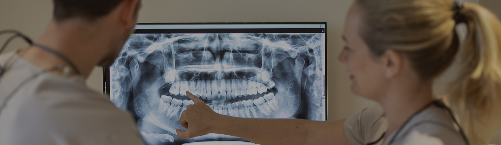
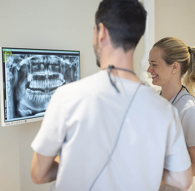
A panoramic X-ray shows all teeth, jaws and maxillary structures in one image. This quick exam is often used for a full dental check-up, orthodontic planning or detecting hidden caries.
3D scanner (Cone beam CT)
3D dental scanning provides high-precision imaging with a low X-ray dose. It is essential before implants, complex surgery or precise endodontic canal identification.
Intraoral radiographs
Intraoral radiography focuses on one or more teeth for detailed views — useful for caries, infection, or checking a filling or crown.
Frequently asked questions
What is the difference between a 3D scan and a panoramic radiograph?
A panoramic radiograph gives an overall view of teeth and jaw, while 3D imaging shows bone and dental structures in three dimensions — useful for implants, precise endodontics or fracture assessment.
Are dental radiology exams painful?
No. They are quick, painless and safe, with low X-ray doses.
When is dental radiology needed?
A dentist may request imaging during a check-up, for unexplained pain, frequent caries, suspected fractures, or before orthodontic or implant treatment.